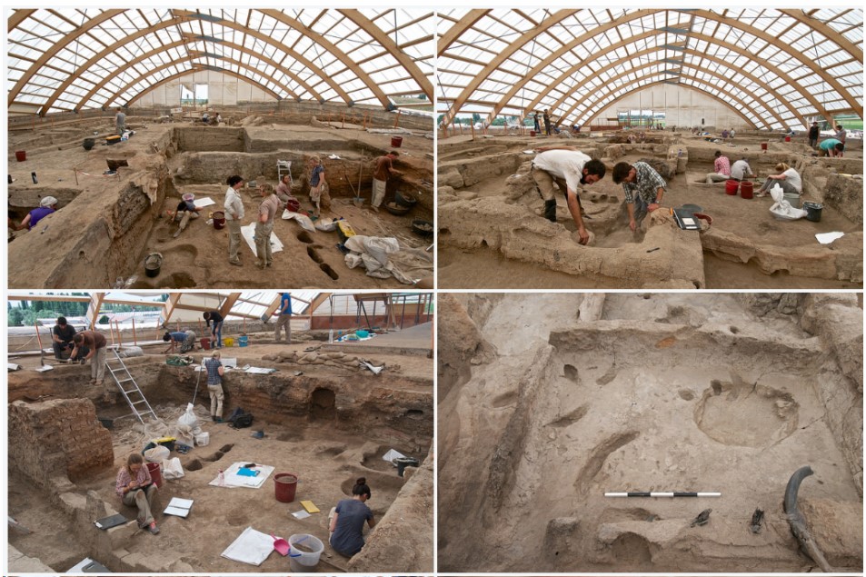
The Neolithic site of Çatalhöyük, first discovered in the late 1950s, is one of the most significant archaeological sites of its period due to its size, population density, and the wealth of artifacts uncovered. Initial excavations were carried out by James Mellaart in the early 1960s, and since then, an international team of archaeologists, led by Ian Hodder, has conducted new excavations and research. These efforts aim to uncover fresh insights into the lives of the population that inhabited this early agricultural settlement. Although not a primary source itself, this field report offers access to information derived from the primary sources unearthed at the site, providing a deeper understanding of Neolithic lifeways.
As you read the field report selection below, consider the following questions:
- What can we learn about the residents of Çatalhöyük by studying dimorphism?
- What does the evidence suggest about the stature and how long residents at Çatalhöyük lived?
- What did the diet of these residents consist of?
- What appears to have been the cause of mortality rates in Çatalhöyük?
Complete the Primary Source Analysis Form when finished.
Source origin: The Human Remains, Theya Molleson and Peter Andrews, Çatalhöyük , last accessed December 12, 2014,
http://www.catalhoyuk.com/archive_reports/1997/ar97_12.html
Summary
A total of at least 64 individuals must have been buried within Room 1. This number includes four neonates in the Fill. With 13 infants under two years and 15 sub adolescent juveniles more than half the sample is of immature individuals. Old adults, of which there are 11 are well represented.
The proportion of immature to adult skeletons is very high but is compatible with an expanding population based on an extended family (or possibly a polygamous family). If each of the areas, NW Platform, 71 and 110 was used by a different nuclear family within the extended family unit this could explain the distribution. The contemporaneous use of the three areas samples sibling (brothers) families at different stages. Thus the youngest family is buried in on the north west plat-form (B38) and their children who died in infancy are buried there in phase I; surviving children that subsequently died in phase II (B36) and phase III (B35).
We can postulate that the family that used area 71 for burial (B30, 40) was already older in phase I than the B38 family and most of the children aged five or more. B31 could contain the dead children of another sibling.
In each case B38, 30, 40, 31 the last burials include an adult female or male; the death of a spouse that ended the family unit. The surviving spouses could have been buried in phase II with the east platform group.
The above describes the demographic pattern of Room 1, its generality can only be confirmed by future work. If the pattern is real there must be a decision to occupy a Building possibly at a point in the segmentation of a family towards the development of a new extended family (cf. Bedouin family structure). The death of the senior member or over-population would mark the end of the life of one extended family, which is the end of the phase of a particular Building.
Evidence for relationships
Three instances of supracondylar fossa of the humerus were noted in individuals buried at different times in area 110. This provides reasonably strong evidence that individuals buried in this area were related and that they in turn were related to 2527 an adult female, who also displays this character and was buried in the Fill before Room I was fully inhabited. Enamel defects noted in two or three cases from area 71 may also indicate related individuals as well as cases of hypodontia or dental reduction.
Spondylothesis, a failure of the last lumbar or first sacral vertebra to unite, has a genetic predisposition in its etiology, and there are two cases of this.
Stature
The people had relatively long forearms and lower legs, a general finding for neolithic samples. Thus, in calculating stature, the formulae of Trotter and Gleser (1952, 1958) given in Brothwell (1981) for negroes were found to give the most consistent results, and have been used here to evaluate heights of females and males. Owing to the fragmented nature of most of the bones few estimates have been possible. The height given in the following Table is that derived from the most reliable bone, where possible a leg bone, rather than an average of different estimates (See Trotter and Gleser 1958, Brothwell 1981).
Dimorphism
Determination of Sex and sexual dimorphism in skeletal material Sex is determined from the manifestation of secondary sex characters which are developed to different degrees in different populations. The characteristics of the pelvis, sacrum and skull are the most distinctive of females; those of the skull, pelvis and sacrum of males. In general, males are more robust than females and measures of robusticity involving two or more diameters may be valuable in determining sex. Such measures should be established for each population since size and therefore robusticity varies between populations. the robusticity of the lower canine can be particularly diagnostic in homogeneous samples, but is of limited value in mixed or
heterogeneous samples.
The use of absolute measurements to evaluate sex is to be avoided. It polarises the sexes, incorrectly attributing large women as males and small men as females. The size range given in many texts for attributing sex (Bass, Standards) will not necessarily apply to the sample under study, which may derive from a population that was taller or shorter than the reference; this being genetically and geographically determined.
Size, especially of males, may be an indicator of environmental conditions and nutritional health. Size of females may relate to envi-ronmental conditions and correlate with age of reproduction. Sexual dimorphism, the difference in size between the two sexes, can be particularly informative of the social structure of the population. In a uniform homogeneous sample there may be distinct differences between the sexes, particularly marked in late growing bones – bones of the jaw, foot, hands, clavicle, patella, and in measures of robusticity of these bones and cortical thickness of the long bones. It should be remembered that robusticity is also a measure of work load/force and may differentiate task specialization.
It is not good practice to use dimensions to infer age of juveniles. This is most reliably done by reference to dental development. The growth achievement of a child can be assessed by comparison of size with dental development. This can give information as to health and genetics. But for demographic purposes, with fragmented material, it may be necessary to resort to the use of dimensions to infer age of immature individuals where the dentition is lacking.
Posture and activity related bone morphology
It seems that a number of different postures were used habitually to rest or to carry out specific tasks: squatting on the heels, squatting or kneeling on toes, both energy efficient (Huard and Montaigne 197x), sitting cross legged, squatting both legs to one side, squatting knees together heels to buttocks, squatting weight on one foot purchase on the other.
Many of these postures may be idiosyncratic, others may be best suited to specific tasks. Pounding ochre with a pestle and mortar would be ergonomically most efficient if the mortar is held between the thighs and the pestle driven from the centre of gravity about the shoulders. Grinding of grain on a saddle quern is best undertaken from a kneeling position with the toes curled under to provide ‘push-off’ for the forward drive. Overshooting the quern and injury to the proximal articulation of the big toe was avoided by placing the quern on a plinth (see Mellaart Anatolian Studies 1962).
One, as yet unidentified, task led to injuries to the thumb. Osteoarthritic changes to the first metacarpal and trapezium of both hands is associated with morphological evidence for squatting on the toes, thighs spread apart.
Handed tasks, leading to arm asymmetry would include wall and floor plastering. These seem to have been onerous tasks especially when old plaster was re-used since it had to be thoroughly broken up (Wendy Mathews has evidence for this from her floor sections).
Diet
In comparison to other Neolithic sites wear on the teeth is very little and even old individuals do not have advanced dental abrasion. This suggests a diet of soft foods, such as those the remains of which have been found in the rooms, including lentils, peas, and acorns. Additionally tubers of water reeds, Scirpus, could have been consumed. Wheat, if eaten must have been in the form of ‘burgul’, not bread which has to be masticated and is abrasive.
There are few cases of dental caries, indicating that refined starch-es, cooked cereals, probably were not available. The few caries include several on the occlusal surface – a phenomenon related to the low abrasion rate.
Periodontal disease is uncommon and lateral abscesses were noted in only two individuals. The food seems to have been consumed in a self cleansing form – large particles and non-glutenous – fruit, nuts, lentils, meat. The generally low levels of calculus fit this impression that food was self cleansing.
A number of individuals with crowding of the anterior teeth would have developed this condition as a consequence of the generally soft food. Generally though there is a surprising lack of dental crowding given the presumed soft nature of the diet.
The diet appears to have been adequate and there are no cases of deficiency disease, although cortical thickness in some children was very thin. General undernutrition is not easy to detect except through evidence for failure to attain expected height at a given (dental) age.
Cause of death
The very high proportion of juveniles in the sample implies a high mortality rate even for young infants, presumably still being suckled.
Epidemics of infectious disease are a possibility, wiping out whole families or returning year after year. Plague, malaria, enteric dysentery are possibilities. The habitual cleanliness of the house would have controlled infection.
The large male, 1466, buried without a head, appears to have been hanged in such a way that he was decapitated, probably before death but possibly after death. It is important to note that the first cervical vertebra and the odontoid peg of the second cervical are missing, while the hyoid and one branch of the cricoid are present. The remaining cervical vertebrae are in full articulation.